Fluorescence microscopy is a microscopic optical observation technique that uses a specific wavelength of excitation light to illuminate an object to produce fluorescence for microscopic examination. It has been used for more than 100 years. It is widely used in biomedical applications. Most laboratories are equipped with high-end or conventional microscopic imaging systems. Fluorescence microscopy is used to study the absorption, transportation, distribution and localization of chemical substances in cells. Some substances in cells, such as chlorophyll, can fluoresce when exposed to ultraviolet light; others can not fluoresce themselves, but if they are stained with fluorescent dyes or fluorescent antibodies, they can also fluoresce when exposed to ultraviolet light. Fluorescence microscopy is One of the tools for qualitative and quantitative research on such substances.
In recent years, various new fluorescent dyes and various specialized fluorescent probes have emerged, such as fluorescent in situ hybridization (FISH) related kits. There are more than ten kinds of probes on the market. Multi, Vysis mFISH Probe Kit (FITC, CY5, TxR, DEAC, SP-Gold), Abbott-FISH Probe Kit (Spectrum Blue, Spectrum Aqua, Spectrum Green, Spectrum Gold, Spectrum Orange), Zytovision FISH (ZyBlue) , DAPI, ZyGreen), Cytocell FISH probe kit, the spectral range of the dye is different, how to configure the appropriate fluorescence microscope to obtain the best fluorescence detection becomes a more complicated problem. Guangzhou Keshi Special Science Instrument Co., Ltd. has been committed to providing customers with customized fluorescence microscope imaging system, based on customer feedback and our own work practices to briefly summarize how to choose the right source to obtain high-quality fluorescence.

A brief principle of fluorescence microscope
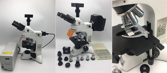
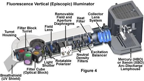
The basic structure of a fluorescence microscope is usually composed of an ordinary optical microscope plus some accessories such as a fluorescent light source, an excitation filter, a two-color beam separator, and a blocking filter. The fluorescent light source used at the same time is generally an ultra-high pressure mercury lamp, which can emit light of various wavelengths, and each fluorescent substance has a wavelength of excitation light which produces the strongest fluorescence, and an excitation filter is usually added, usually ultraviolet. The purple, blue, and green excitation filters only need to transmit a certain wavelength of excitation light to the specimen and absorb other light.

two. Factors affecting the imaging effect of fluorescence microscopy
There are many factors affecting the fluorescence microscope imaging, including: preparation of fluorescent samples, selection of fluorescent light sources, design of filter discs, and combination of fluorescent filters (excitation filter, emission filter, dichroic filter) , bandwidth selection), the type of microscope objective, the choice of microscope camera system and other issues. Fluorescence excitation, filtering, imaging, quenching, adjustment of fluorescence experiments is how to coordinate the above factors to get the best results, one of the important factors affecting the imaging effect is the fluorescent light source.

three. Fluorescence microscope light source types and introduction
Common light sources for fluorescent microscopes are usually white light sources: high-pressure mercury lamps, xenon lamps, metal halide lamps and LED white light sources, and high-end LED single-wavelength sources.
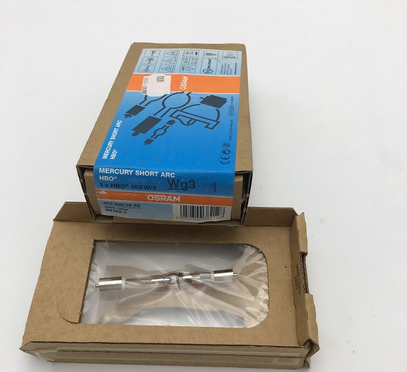
1. Mercury lamp <br> Ultra-high pressure mercury lamp (50-200W), which is made of quartz glass with a spherical shape in the middle and filled with a certain amount of mercury. It is discharged between two electrodes during operation, causing mercury to evaporate inside the ball. The air pressure rises rapidly, and when the mercury is completely evaporated, it can reach 50 to 70 standard atmospheric pressures, and the process generally takes about 5 to 15 minutes. The luminescence of an ultra-high pressure mercury lamp is the result of the discharge of light during the continuous dissociation and reduction of mercury molecules by the discharge between electrodes. It emits strong ultraviolet and blue-violet light, which is sufficient to excite various types of fluorescent substances, and is therefore commonly used for fluorescence microscopy. Recommended products: Osram 50W, 103W, 200W mercury lamp, suitable for high-end fluorescence microscopes of major manufacturers; generally 200 hours life.

2, xenon lamp <br> Xenon lamp and mercury lamp as a white light source, can provide a range of wavelengths from ultraviolet to near infrared, but they have different excitation spectra. Mercury lamps are concentrated near the near ultraviolet, blue, and green light. This high excitation energy at the peak ensures a bright fluorescent signal, but the phototoxicity is strong. Therefore, HBO is used to image fixed samples or weak fluorescence. In contrast, xenon lamp source excitation is relatively flat and can be used for comparison of intensity between different wavelengths, for example for calcium ion concentration measurement. The strong excitation of the xenon lamp is in the near infrared 800-1000 nm.
Features of XBO over HBO:
1) More uniform spectral intensity
2) Strong spectral intensity in infrared and mid-infrared
3) Stronger energy and easier access to the objective lens. Such products: XBO75W, 100W xenon lamp; life expectancy is 400 hours, 1200 hours.
3. Metal Halogen Lamps <br> A light source widely used by major microscope brands. It is similar to the excitation spectrum of mercury lamps, but it is connected to the microscope by fiber, which can dissipate heat and last longer. The fiber is connected from the light source to the microscope, which can greatly reduce the heat generated by the light. Another advantage of metal halide lamps for imaging live cells is the built-in intensity design that makes it easy to adjust the excitation light. You can also get better excitation by choosing a direct connection. For example: EXCELITAS X-cite light source, 120W, suitable for high-end fluorescence microscopes of major manufacturers; life expectancy of more than 2000 hours. Guangzhou Keshi Special Science Instrument Co., Ltd. is the authorized agent of this company. Price concessions, fast delivery.
External Aluminum Foil Reflective Series
External Aluminum Foil Reflective Series,Sunshade Net with Grommets,Wind Resistant Outdoor Shade Net,Insulation Sun Shade Curtain
Changzhou Green Nets Co.,Ltd. , https://www.czglnets.com